Human Anatomy Female Abdomen - Female Anatomy Diagram High Resolution Stock Photography ... / Human body anatomy yoga anatomy human anatomy and physiology muscle anatomy knee muscles anatomy anatomy study.
Human Anatomy Female Abdomen - Female Anatomy Diagram High Resolution Stock Photography ... / Human body anatomy yoga anatomy human anatomy and physiology muscle anatomy knee muscles anatomy anatomy study.. A regional study of human structure. National library of medicine was used as the basis to build an. Anatomy of female abdomen, download this wallpaper for free in hd resolution. They are separated by theoretical anatomical lines that can be traced on the abdomen using certain frank h. Exemplary model of the female abdomen and pelvis.
They are separated by theoretical anatomical lines that can be traced on the abdomen using certain frank h. Blood vessels, lymphatic drainage and nerves of the abdomen. Thus, the right side of the image is the patient's left. The abdomen (colloquially called the belly, tummy, midriff or stomach) is the part of the body between the thorax (chest) and pelvis, in humans and in other vertebrates. Two ways of dividing the abdomen into regions.
Abdominal Cavity, kidneys model | Medical anatomy, Medical ... from i.pinimg.com The human abdomen is that part in the front of our body between the chest and the waist line. Anatomy of female abdomen, download this wallpaper for free in hd resolution. Human body anatomy yoga anatomy human anatomy and physiology muscle anatomy knee muscles anatomy anatomy study. Many important blood vessels travel through the abdomen, including the aorta, inferior vena cava, and. Exemplary model of the female abdomen and pelvis. In the female the peritoneum is not a closed sac, since the free ends of the uterine tubes open directly into the peritoneal cavity. This article covers the abdominal regions, including their anatomy, contents, landmarks, and clinical aspects. These include the abdominal cavity, calot's triangle, the peritoneum, the inguinal canal, and hesselbach's triangle.
To delineate organ outlines and.
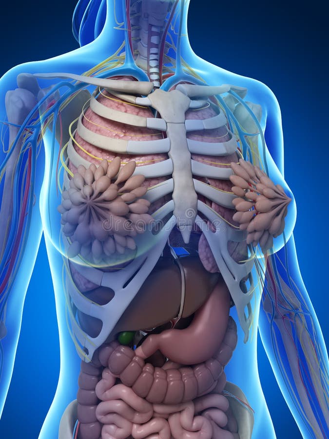
Many important blood vessels travel through the abdomen, including the aorta, inferior vena cava, and. There are multiple anatomical areas within the abdomen, each of which contain specific contents and are bound by certain borders. Abdominal wall & cavity the abdomen is the part of the trunk inferior to the thorax. Let's take a close look at this very important part of our anatomy and thus improve our understanding of causes of abdominal pain. The human abdomen is that part in the front of our body between the chest and the waist line. In the female the peritoneum is not a closed sac, since the free ends of the uterine tubes open directly into the peritoneal cavity. Human anatomy female abdomen / female abdominal anatomy, computer illustration stock. National library of medicine was used as the basis to build an. Anatomy of liver the liver is a reddish brown organ with four lobes of unequal size and shape. Human female (vhf) for laparoscopic surgery training. There are many pics about muscles human body diagram out there. It is of an oval shape, the extremities of the oval being directed upward and downward. If you want to learn how to read ct scans of the abdomen and pelvis proficiently, this video is an excellent starting point.
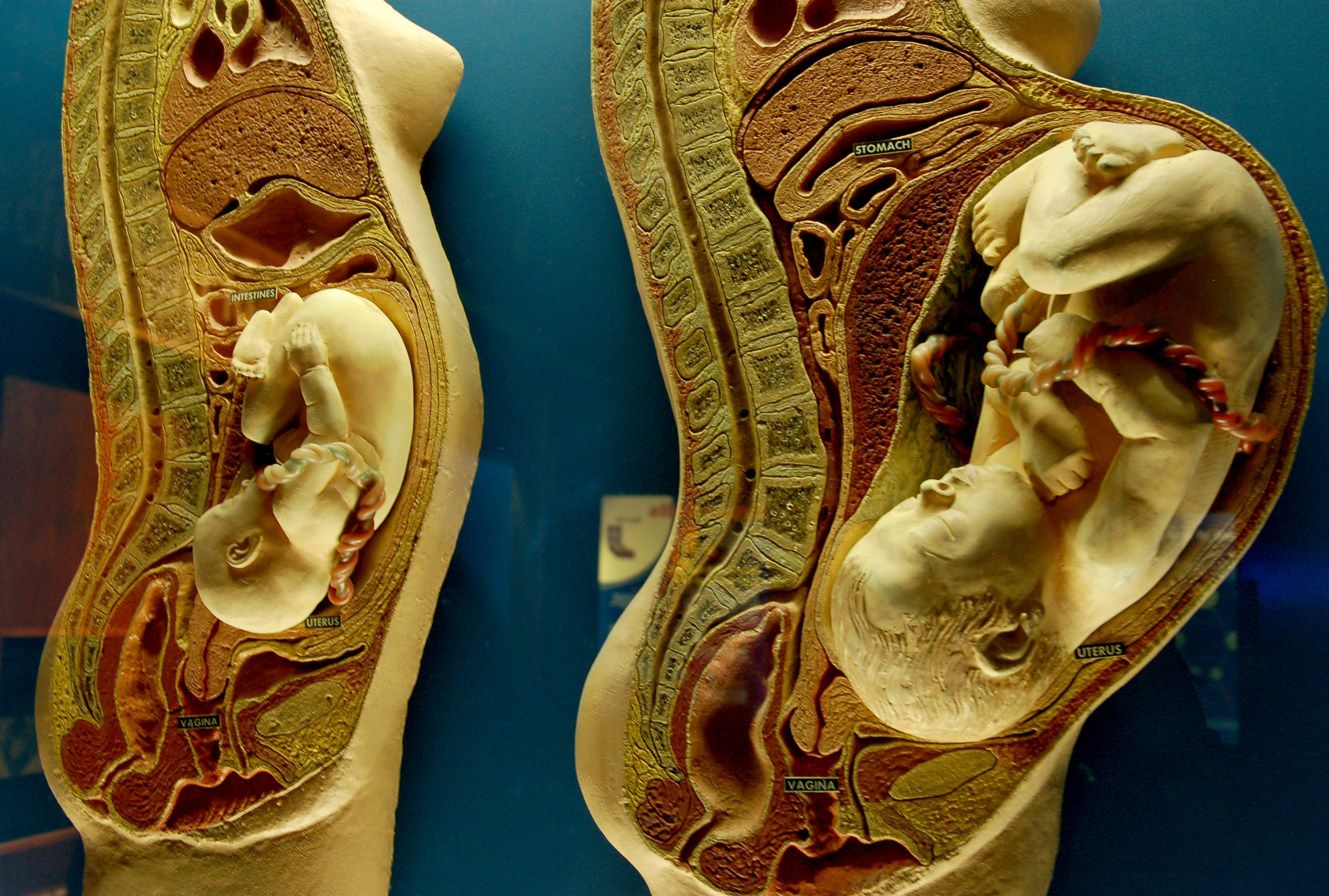
Abdominal wall & cavity the abdomen is the part of the trunk inferior to the thorax. To delineate organ outlines and. Diseases affecting any of these organs could result in abdominal pain. The standard position of the uterus is anteverted and anteflexed. Exemplary model of the female abdomen and pelvis.
Female Anatomy Stock Photography - Image: 30725622 from thumbs.dreamstime.com Let's take a close look at this very important part of our anatomy and thus improve our understanding of causes of abdominal pain. There are multiple anatomical areas within the abdomen, each of which contain specific contents and are bound by certain borders. Anatomy of liver the liver is a reddish brown organ with four lobes of unequal size and shape. They are separated by frank h. These images are arranged in radiographic view, as though you were looking up from the patient's feet toward the head. • we're going to take apart a plastic anatomy model and see what we. The bones of the abdomen are made up of the lumbar. its musculomembranous walls surround a large cavity (the 28.
Female abdominal thoracic anatomy medical illustration.
The video covers the most. The standard position of the uterus is anteverted and anteflexed. The bones of the abdomen are made up of the lumbar. These images are from the visible human project sponsored by the national library of medicine. If you want to learn how to read ct scans of the abdomen and pelvis proficiently, this video is an excellent starting point. Diseases affecting any of these organs could result in abdominal pain. Both pass through a skeletal muscle (voluntary) sphincter in the urogenital. This hd wallpaper anatomy of female abdomen has viewed by 846 users. its musculomembranous walls surround a large cavity (the 28. The four anatomical regions of the abdomen are known as quadrants. The liver, stomach, large intestines, rectum, uterus, vaginal. Exemplary model of the female abdomen and pelvis. Related posts of anatomy of the abdomen female muscles human body diagram.
Female anatomy, early 17th c wellcome l0011866.jpg 1,178 × 1,707; The abdomen is the largest cavity in the body. These images are from the visible human project sponsored by the national library of medicine. Human female (vhf) for laparoscopic surgery training. Human anatomy lesson 15 abdomen.
Pregnant woman anatomy - BabyCenter from farm4.static.flickr.com The bones of the abdomen are made up of the lumbar. • we're going to take apart a plastic anatomy model and see what we. Image from marieb et al., human anatomy, 7th edition, pearson education, 2014. This full color custom medical exhibit features an anterior and sagittal view of the normal anatomy of the female reproductive system, an enlarged anterior view of the left fallopian tube and ovary is included. In the female the peritoneum is not a closed sac, since the free ends of the uterine tubes open directly into the peritoneal cavity. Thus, the right side of the image is the patient's left. Blood vessels, lymphatic drainage and nerves of the abdomen. Don't forget to share this picture with others via facebook, twitter, pinterest or other social medias!
National library of medicine was used as the basis to build an.
Organs shown and labeled are: This full color custom medical exhibit features an anterior and sagittal view of the normal anatomy of the female reproductive system, an enlarged anterior view of the left fallopian tube and ovary is included. Image from marieb et al., human anatomy, 7th edition, pearson education, 2014. The standard position of the uterus is anteverted and anteflexed. National library of medicine was used as the basis to build an. A regional study of human structure. Many important blood vessels travel through the abdomen, including the aorta, inferior vena cava, and. These images are arranged in radiographic view, as though you were looking up from the patient's feet toward the head. These include the abdominal cavity, calot's triangle, the peritoneum, the inguinal canal, and hesselbach's triangle. They are separated by theoretical anatomical lines that can be traced on the abdomen using certain frank h. The video covers the most. Female reproductive system anatomy digestive system anatomy human digestive system human body systems cardio rectus abdominis muscle abdominal schematic cross section of abdomen at middle t12 anatomy liver, falciform ligament, superior epigastric vessels, transversalis fascia. Blood vessels, lymphatic drainage and nerves of the abdomen.


0 Komentar